THIS IS HOW IT HAPPENED
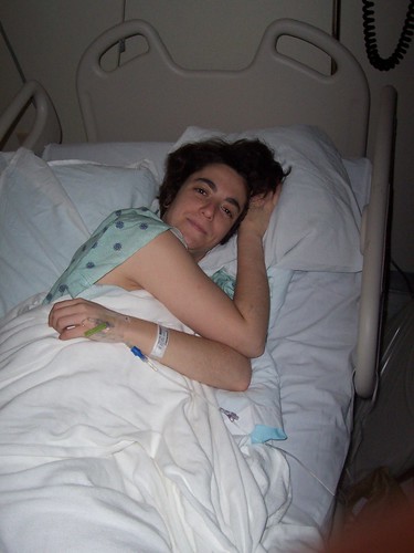
On Sunday, November 2, I started to feel more tired than usual. After work Monday and Tuesday, I came home and went straight to bed. Wednesday at work I could not walk to The Landing without sitting down several times (it is only about 50 feet between buildings). After vomiting that day, my coworkers kicked me out of the building and sent me to the doctors.
My doctor treated me for asthma and sent me for blood tests and a chest xray, which I had done the next day. Meanwhile, I slept all day Thursday and Friday, barely able to walk to the bathroom without becoming extremely fatigued. By Friday night, I was frustrated and not convinced that the asthma diagnosis was correct. My friends Beth and Lee brought me to the Emergency Room at St. Vincent's Hospital.
I was admitted to the hospital when the doctor ran an EKG and found some irregularities in my heartbeat. I was immediately hooked up to a heart monitor which I wore for the remainder of my stay.

On Saturday morning, I met my cardiologist, Dr. Jonathan Constantin. I nicknamed him Dr. Bling Bling because he is quite handsome and a snappy dresser. I later learned that others called him Dr. Gigalo. Regardless, he proved to be a very compassionate and informed doctor.
The first test I was given was an echocardiogram.
The first test I was given was an echocardiogram.

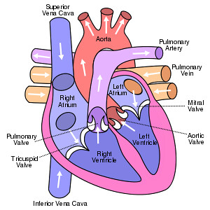
What is an Echocardiogram?
An echocardiogram is a test in which ultrasound is used to examine the heart. The equipment is far superior to that used by fishermen. In addition to providing single-dimension images, known as M-mode echo that allows accurate measurement of the heart chambers, the echocardiogram also offers far more sophisticated and advanced imaging. This is known as two- dimensional (2-D) Echo and is capable of displaying a cross-sectional "slice" of the beating heart, including the chambers, valves and the major blood vessels that exit from the left and right ventricle. Above is an example.
The test showed that I had an enlarged left ventricle which was only ejecting oxygen-rich blood to my body at 10%. Average ejection fraction is 50%. Ejection fraction (Ef) is the fraction of blood pumped out of a ventricle with each heart beat.
Finding out that my heart was pumping out very little blood was very scary. I was relieved to finally know what was wrong with me. It just didn't seem like it should be happening to me. Maybe they read the wrong test. I mean, I am 33 years old. Even my doctor said I was "special". These are problems that older people deal with. Heart failure was not something I thought I had to worry about.

Dr. Constantin explained that cardiomyopathy can be caused by a virus that settles in the heart. Searching for the virus would be a waste of time because it would not alter the treatment at all. Dilated cardiomyopathy can be inherited. It also can be caused by certain diseases, conditions, and substances, including:
· Coronary artery disease and heart attacks
· Infections, especially viral infections that effect the heart muscle
· Alcohol, especially when a person has a poor diet
· Complications during pregnancy
· Certain toxins, such as cobalt
· Drugs, such as cocaine, amphetamines, and some cancer medicines
· Diseases such as diabetes and thyroid disease
The next test I had was a heart catheterization to make sure I did not have any blockages.
What is cardiac catheterization?
What is cardiac catheterization?

This is a procedure to examine blood flow to the heart and test how well the heart is pumping. A doctor inserts a thin plastic tube (catheter) (KATH'eh-ter) into an artery or vein in the arm or leg. From there it can be advanced into the chambers of the heart or into the coronary arteries. This test can measure blood pressure within the heart and how much oxygen is in the blood. It's also used to get information about the pumping ability of the heart muscle.
I was given a sedative and a local anesthetic during the procedure. Although I could have watched everything, I fell asleep. I awoke to a nurse holding pressure on my thigh . That was strange. I had to lay flat without moving for four hours following the procedure. After it was all done, I only had two small pinholes on my upper right thigh. I am pleased to report that no blockages were found.
With these tests finished, Dr. Constantin was able to determine the best course of treatment. When he mentioned changes in lifestyle, diet, habits, and the possible need for a defibrillator and perhaps a heart transplant, I have to admit that I was shocked, angry, and sad. My parents shared my range of emotions.

While in the hospital, my sister Anne Marie was a constant consultant as a former respiratory therapist. She helped us figure out which questions to ask, what to expect, and helped put everything in perspective.
After all of this, on day seven of my hospital stay, I was subjected to the worst test ever - an MRI. I developed a pilonidal cyst at the base of my spine after about 5 days in the hospital. When I complained of the soreness, everyone thought it was a bed sore. I had no bump, no redness, and no pain when I entered the hospital. I was given an egg crate to sit on and some cream. Finally, after a large, pinkie shaped/sized sore developed, the general medicine doctor, Dr. Doan, or as I prefer to call him, Dr. Duh, ordered an MRI. "An MRI for my butt?" I asked.
What is a pilonidal cyst?

A pilonidal cyst is a unique kind of abscess that occurs in or above the crease of the buttocks. Pilonidal cysts often begin as tiny areas of infection in the base of the area of skin from which hair grows (the hair follicle). With irritation from direct pressure, over time the inflamed area enlarges to become a firm, painful, tender nodule making it difficult to sit without discomfort. These frequently form after long trips that involve prolonged sitting.
MRIs SUCK!!!
They tape you down, stick you in a tube (mine was smaller than the one in this picture), put ear plugs in your ears, turn on a wicked loud roaring noise, and leave you there FOREVER. Then, they pull you out, stick "contrast" in your arm, and put you back in again.
MRIs SUCK!!!
They tape you down, stick you in a tube (mine was smaller than the one in this picture), put ear plugs in your ears, turn on a wicked loud roaring noise, and leave you there FOREVER. Then, they pull you out, stick "contrast" in your arm, and put you back in again.

It was AWFUL! I had to lie on my back, where the sore was located - that hurt a lot! And I had to go to the bathroom. By the time it was over, I was squeezing the emergency bell and excited to return to my room.
The MRI revealed that the bump was a pilonidal cyst. That meant surgery to drain and remove it. That meant a few more days in the hospital. My care shifted from my heart condition to a bump on my butt. I went into surgery at 9:30am, was in the operating room by 11:15am, and out of recovery by 12:30pm.
The MRI revealed that the bump was a pilonidal cyst. That meant surgery to drain and remove it. That meant a few more days in the hospital. My care shifted from my heart condition to a bump on my butt. I went into surgery at 9:30am, was in the operating room by 11:15am, and out of recovery by 12:30pm.
I was not able to be anesthetized because my ejection fraction is so low, so I was given a local anesthetic and a sedative. Luckily, I fell asleep again. All I remember is moving onto the operating table, being covered with warm blankets, thinking that there were a lot of people needed to remove a bump, and noticing how cold my exposed tukus felt. When I was roused from my slumber, I thought I was in an episode of Doctor Who. I swore I was in a hospital bed in the Tardis. It seemed very real.

Every day since the surgery, the wound has been cleaned and dressed. A home care nurse comes every few days to take my vitals and to take a look at the incision. Earlier this week, a bit of infection was detected. I went to Solantic for a culture and we are waiting to find out the results. Yeah!
.svg.png)

No comments:
Post a Comment