
I just returned from my cardiologist, lovingly nicknamed “Dr. Bling Bling.”
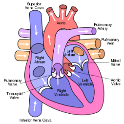
Last week, I had an echo-cardiogram to determine if my heart has improved since I was in the hospital four months ago. The test showed that my heart has strengthened and is returning to its normal shape. My ejection fraction has improved, but not enough to take me out of the danger zone. My ef is up from 10%, but still well below 35%, so I will be seeing a doctor that specializes in defibrillators next week. On Monday, I am scheduled to have a T Wave test.

What’s that?
The T wave alternans (TWA) test uses an electrocardiogram (ECG) measurement of the heart's electrical conduction.

The microvolt T-wave alternans (MTWA) is a non-invasive heart test that can identify patients who are at increased risk of sudden cardiac death. It is most often used in patients who have had myocardial infarctions (heart attacks) or other heart damage to see if they are at high risk of developing a potentially lethal cardiac arrhythmia.
Those who are found to be at high risk would therefore benefit from the placement of a defibrillator device which can stop an arrhythmia and save the patient's life.
For more information on the T-wave test,
visit: http://en.wikipedia.org/wiki/T_wave_alternans

Bottom Line
The results of my echocardiogram alone were not enough to determine whether or not a defibrillator is necessary. The T-wave test will provide additional information to determine this.
My heart is working better than it was four months ago, but still below the minimum acceptable levels.
I will continue to take medication – Coreg was doubled to continue the progress of strengthening the heart.
My heart is still alarmingly weak and I am still at high risk of heart failure.

After the test on Monday and the consultation with the "electrician" (cardiologist specializing in the electronics of the heart) on Wednesday, I will know more about the defibrillator.

Surgery to Implant a Defibrillator (According to hearthelp.com)
Implanting a heart device involves making a small incision, approximately 2 inches, in the upper chest, and guiding leads (thin insulated wires) through a vein and into your heart. It is not an open heart procedure. Patients are generally awake throughout the procedure, and a local anesthesia is typically used.
Your doctor will then connect the lead wires to the implanted heart device and program the device settings. Finally, the heart device will be inserted beneath the skin and the incision in your chest will be closed. Most people stay in the hospital overnight and go home the next day.
Before the procedure you will be given medication to make you sleepy and comfortable. After the implant you may see a slight bulge under your skin where the device is located. The leads are very thin and will not be visible.
You will usually stay in the hospital overnight and receive instructions for home care. You may be asked to restrict movement of the arm near the device for a short period after the implant.


.svg.png)

